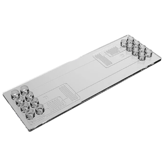
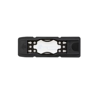
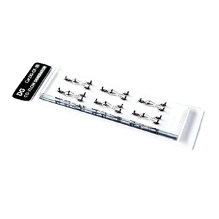
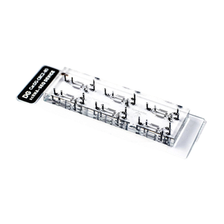
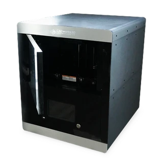
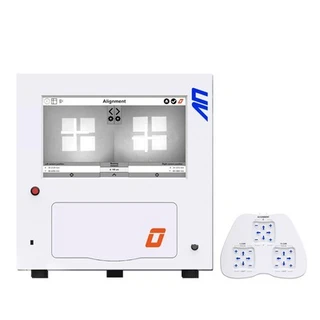
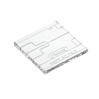
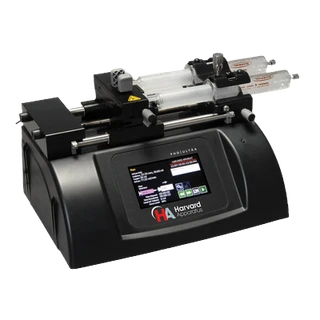
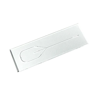
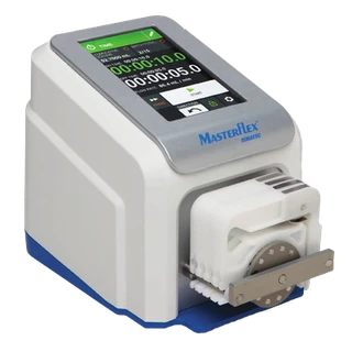
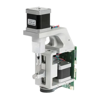
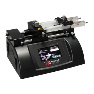
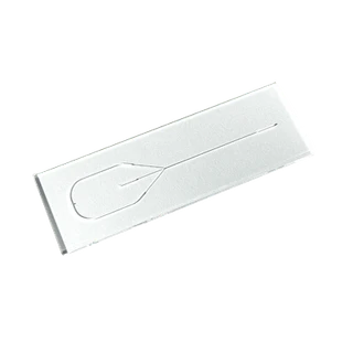
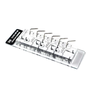
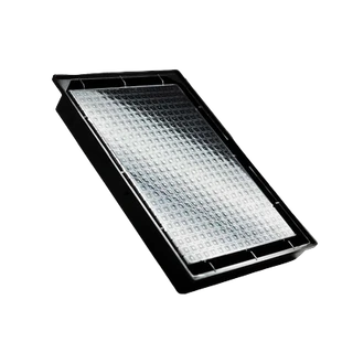
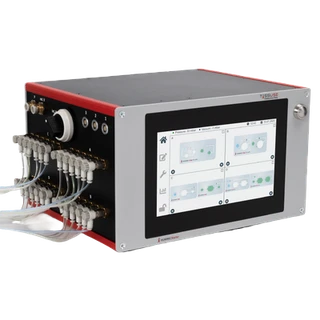
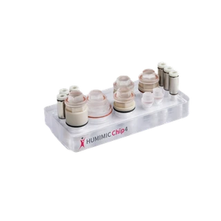
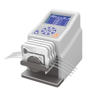
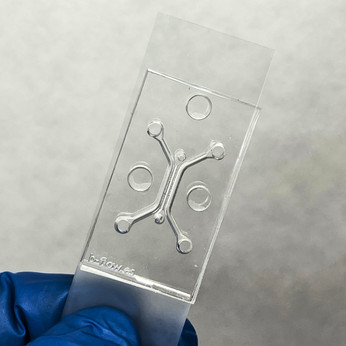
This Organ-on-a-Chip with central chamber and flanked microchannels allows to create 3D tissue models of the brain that accelerate real-time studies of cellular interactions, extravasation and drug delivery. It provides a morphologically and biologically realistic microenvironment that more accurately depicts the in vivo reality.
General
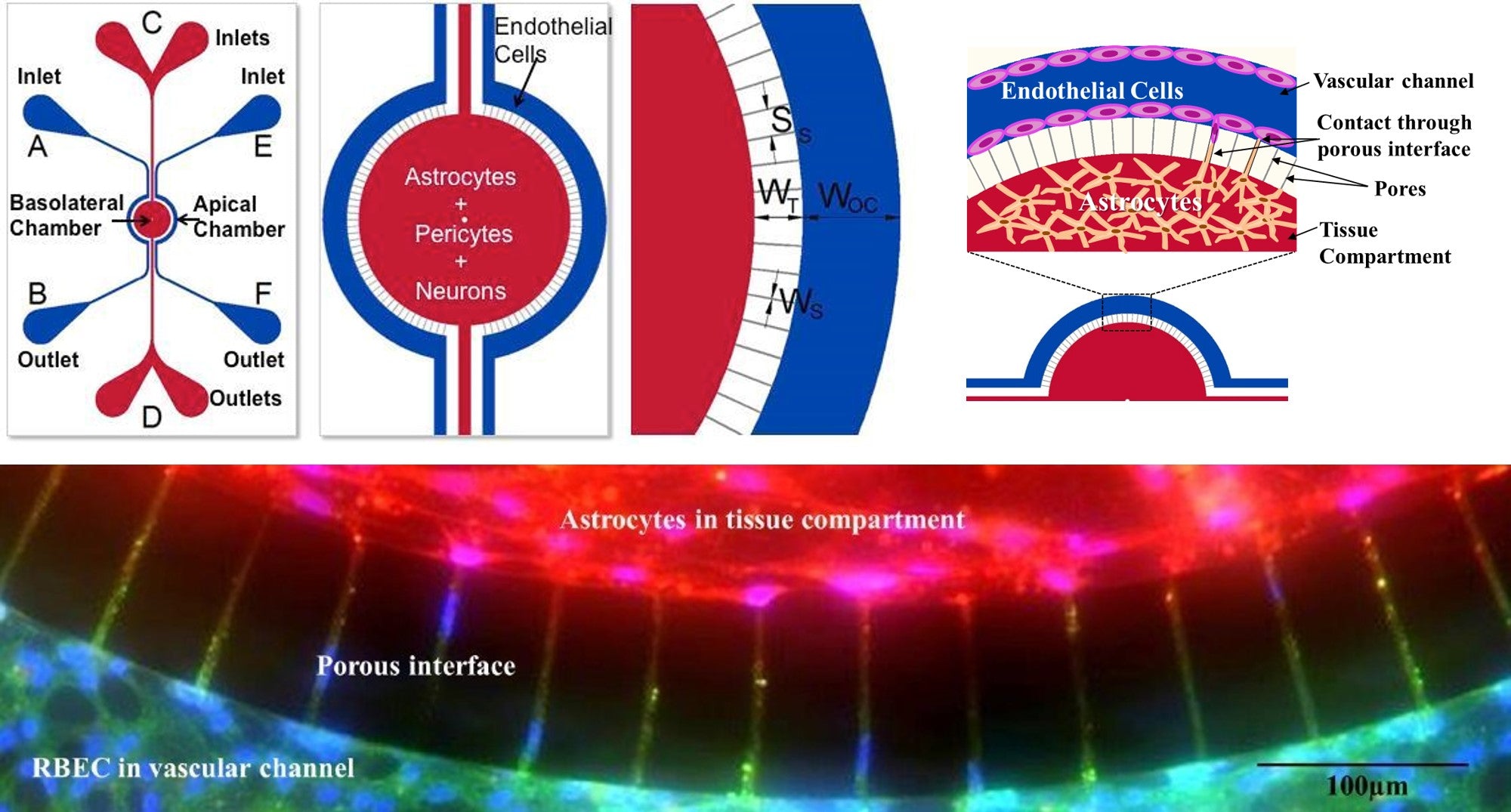
SynBBB is a microfluidic device that allows recreating the complexity of the brain in vivo microenvironment by reproducing a histological section of brain tissue cells in communication with endothelial cells across the blood-brain barrier (BBB). Interactions between brain tissue cells and endothelial cells are easily observable in the SynBBB model through biochemical or electrical analysis.
SynBBB is the only in vitro model of the BBB that allows :
- a precise hemodynamic shear stress in vivo
- real-time observation of drug transport, cellular and barrier functionality
- compatibility with standard analytical instruments
- a robust and easy-to-use protocol
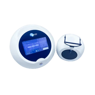
In addition, the IMN2 TEER option available with this model allows you to accurately measure electrical resistance, providing a non-invasive method for real-time monitoring of tight joints. Indeed, the formation of tight junctions between cells (e.g. the blood-brain barrier) can be assessed by measuring changes in electrical resistance in the intercellular space between cells. The SynVivo Cell Impedance Analyzer, used in combination with the SynBBB TEER device, measures electrical impedance (resistance).
Starter Pack - To order the chip only, check our dedicated pages for linear and radial designs (ref SY-102005, SY-102015, SY-108011).
Content
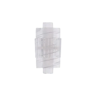
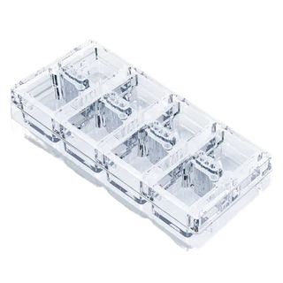
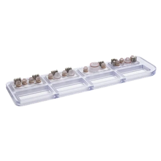
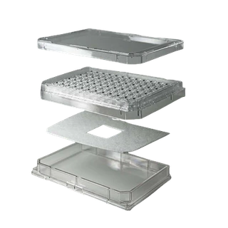
12x SynBBB chip (choice of IMN2 radial, IMN2 linear or IMN2 radial-TEER co-culture chips)
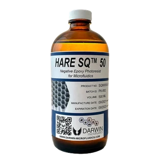
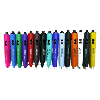
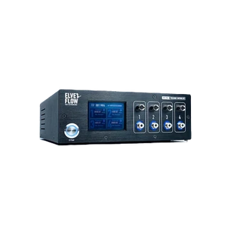
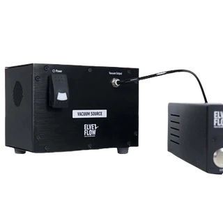
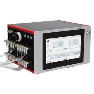
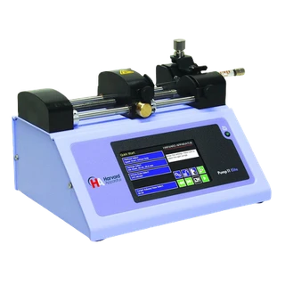
1x Pneumatic priming device (required for priming tubing to remove air)
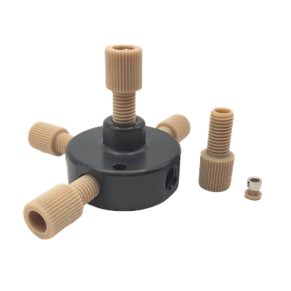
1x Manifold (allows for multiple devices)
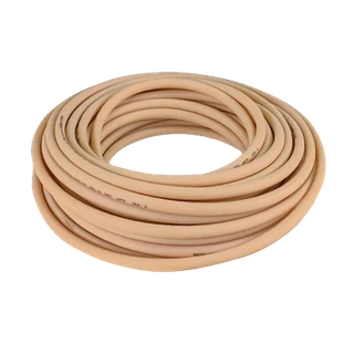
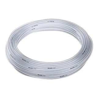
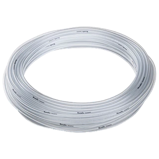
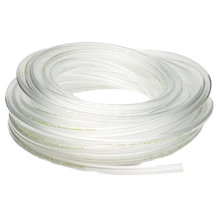
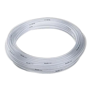
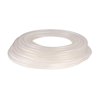
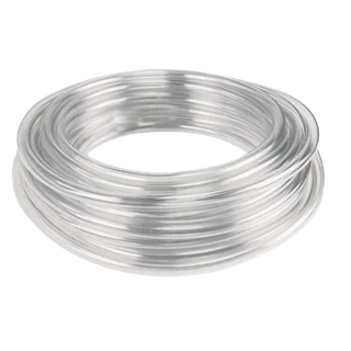
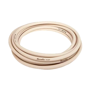
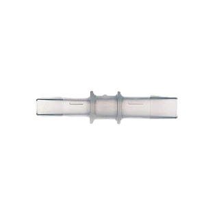
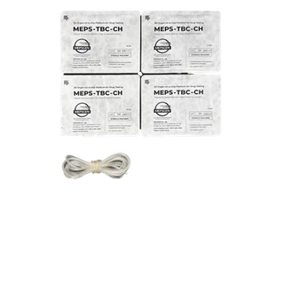
1x Tygon Tubing .02"ID X .06"OD (100 ft)
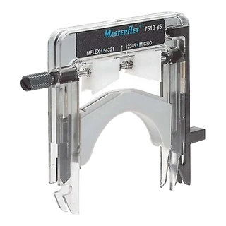
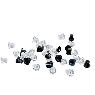
25x Slide Clamps
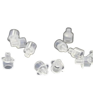
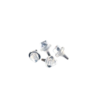
50x Blunt Tip Needles
50x 1mL Syringes with Luer-Lok® Tip
1x Impendence Analyzer required for SynBBB TEER measurements (Only in IMN2 TEER kit)
20x Electrodes (Only in IMN2 TEER kit)
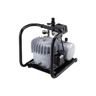
This kit does not include the air pump needed to establish the air-liquid interface.
Specifications
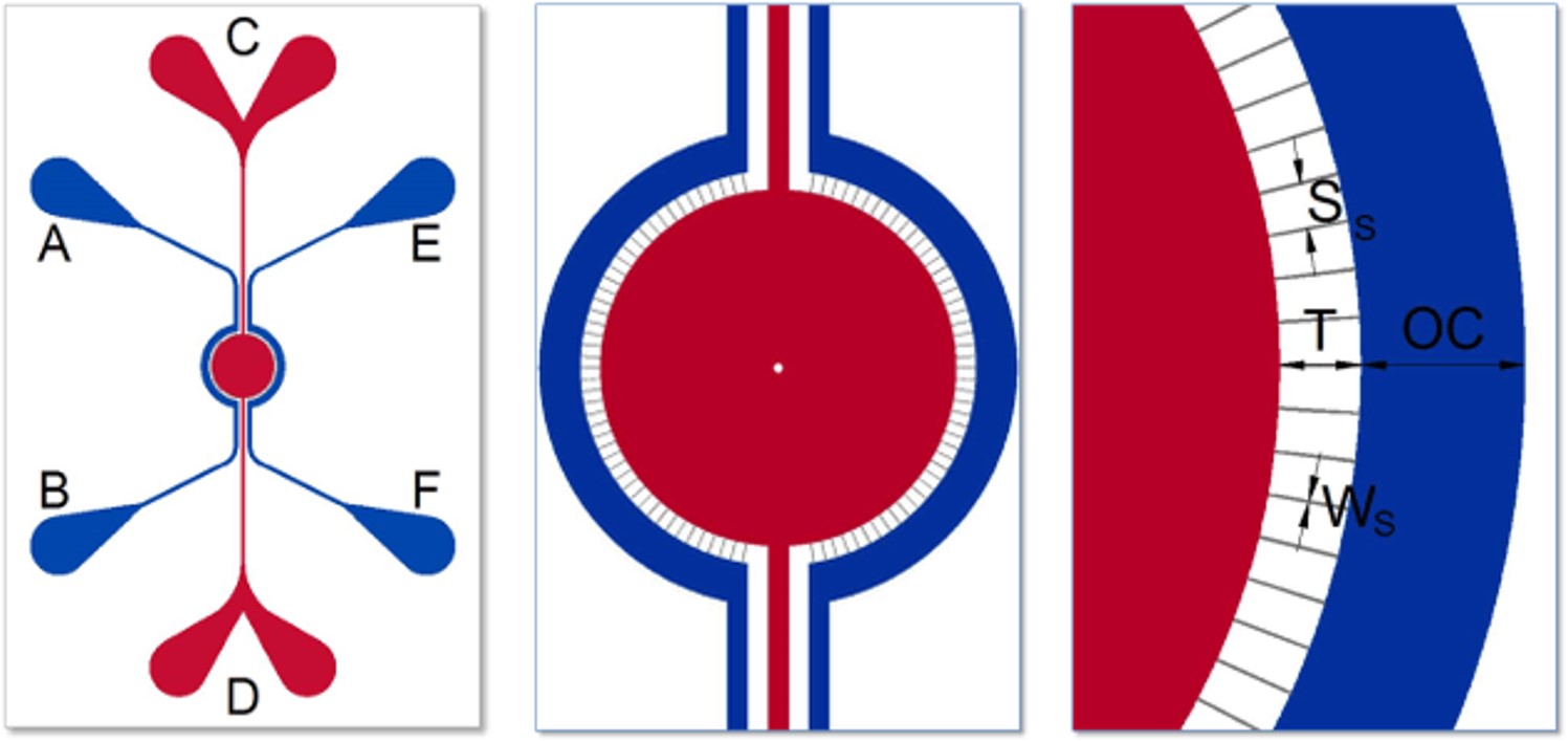
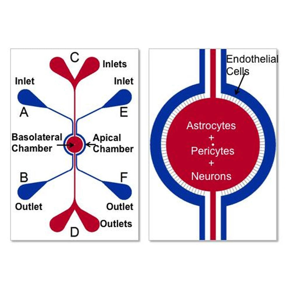
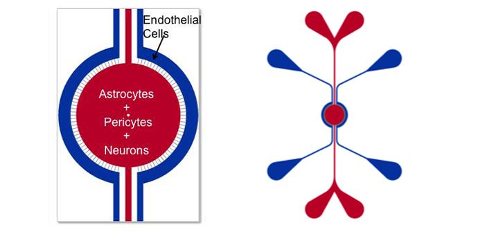
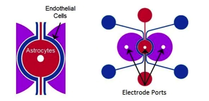
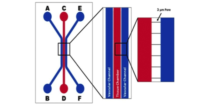
Schematics of the devices used to develop the BBB Model. Apical chamber (outer channels) are for culture of vascular (endothelial cells) while basolateral chamber (central chamber) are for culture of brain tissue cells (astrocytes, pericytes, or neurons). Porous architecture enables communication between the vascular and tissue cells. Outer Channel Width (OC), Travel Width (T), Slit Spacing (SS), Slit Width (WS).
| IMN2 radial |
IMN2 TEER |
IMN2 linear |
 |
 |
 |
|
Outer channel (OC): 200 µm
Tissue chamber (red): 1.8 mm diameter
Slit spacing (SS): 50 μm
Slit width (WS): 3 µm
Travel (T): 50 μm
Chip depth: 100 μm
|
Outer channel (OC): 200 µm
Tissue chamber (red): 1.8 mm diameter
Slit spacing (SS): 50 μm
Slit width (WS): 3 µm
Travel (T): 50 μm
Chip depth: 100 μm
w/ impedance capability
|
Channels width: 200-500-200 µm
Tissue chamber (red): 1.8 mm diameter
Slit spacing (SS): 50 μm
Slit width (WS): 3 µm
Travel: 50 μm
|
Applications
Many co-culture protocols have been developed to establish true vascular monolayers in communication with tissue cells. Human cells grown in these chips retain a biological phenotype similar to that found in the real tissues. Leading researchers have validated that cells grown in these chips more accurately reflect the cell behavior found in vivo compared to cells grown using conventional culture techniques.
The available microfluidic platforms can be used to study cell/particle adhesion and cell-cell or cell-drug interactions and has been extensively validated across neuroscience, oncology, inflammation and toxicology applications.
Unlike well-plate tests performed under static conditions, these chips reproduce the realistic dynamic conditions for the assessment of cell-drug and cell-cell interactions thereby providing an accurate in vitro platform to study and elucidate the mechanisms of success and failure. Compared to in vivo animal studies, they allow real-time visualization and analysis of the assay in a controlled environment.
Under physiological fluid flow conditions it would be possible to study the interactions during:
- Inflammation (leukocyte-endothelium),
- Cancer development (tumor-endothelium),
- Thrombosis (platelet-endothelium),
- Infection (microbial-endothelium)
3D Cancer models
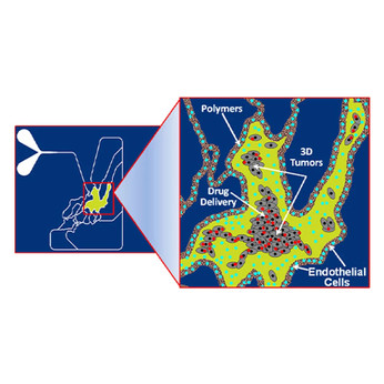
You can create a microfluidic 3D cell-based assay platform for quantitative assessments in a physiologically realistic tumor microenvironment. The system enables real time visualization and analysis of cell-cell and cell-drug interactions encompassing (a) transport across the vessel walls, and (b) delivery to the tumors. There are many areas of oncology research that can benefit by using these models. These include (1) basic research for understanding of the tumor microenvironment (cell viability, proliferation, invasion and tumor-stromal and tumor-endothelium interactions); and (2) drug screening for efficacy, toxicity and penetration.
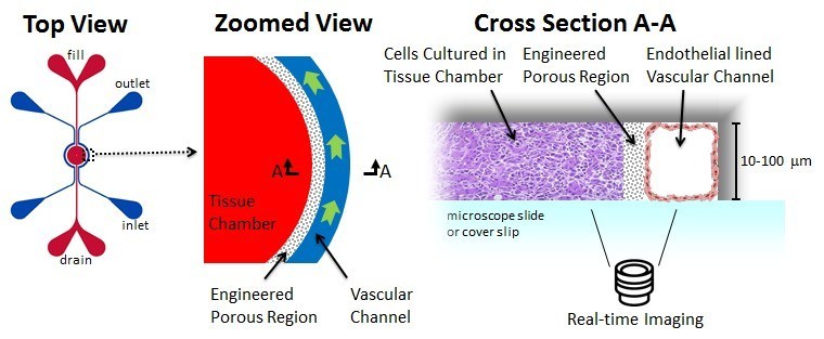
Chips with Design 1 allow to study cancer metastasis. This is a multi-step process that starts with the cancer cells leaving the original tumor site and migrating to distant parts of the body via the bloodstream or the lymphatic system. This process involves complex steps, including breaking of the extracellular matrix by the metastatic tumor cells, escape into the circulatory system, adhesion to the vascular wall at remote locations, followed by migration/invasion into tissue and subsequent proliferation.
Chips with Design 2 are suitable for drug screening. Drugs or delivery systems (nanoparticles, polymers, liposomes, etc.) can be injected via the vascular channel or directly in the tumor chamber under both static and physiological fluid flow conditions and their responses can be observed in real-time mimicking the in vivo conditions.

3D cancer model technical manual
Blood-Brain-Barrier model
The BBB 3D model recreates the in vivo microenvironment by mimicking the histology of brain tissue cells in communication with endothelial cells across the BBB. Shear-induced endothelial cell tight junctions, which cannot be achieved in the Transwell® model, are easily achieved in the this model using physiological fluid flow. Interactions between brain tissue cells and endothelial cells are readily visualized in this assay. Transwell models do not allow real-time visualization of these cellular interactions, which are critical for understanding of the BBB microenvironment.
The Chip with Design 1 - option A (slit barrier) is a highly versatile platform for investigation of:
- Tight junction proteins: Determine the levels of tight junction proteins namely zonula occludens, claudins and occludins which regulate the BBB.
- Transporter proteins: Analyze functionality of transporter proteins (e.g. Pgp) in normal and dysfunctional BBB.
- Drug permeability: Evaluate real-time permeability of therapeutics and small molecules across the endothelium of the BBB.
- Inflammation: Understand the underlying mechanisms of inflammatory responses on the regulation of the BBB.
- Cell migration: Visualize and quantify in real-time migration of immune cells across the BBB.
- Omic changes: Perform genomic, proteomic and metabolic analysis on normal and dysfunctional BBB.
- Neurotoxicity: Analyze toxicity effects of chemical, biological and physical agents on the cells of the BBB.
- Neuro-oncology: Investigate effects of the tumor cells on the BBB.
3D BBB model technical manual
Toxicology model
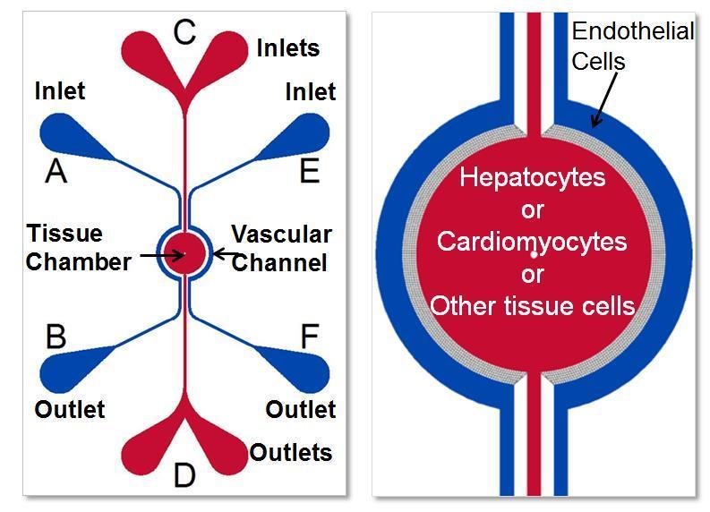
Current in vitro models use 2D monolayers or 3D aggregates of cells under static conditions for studying drug toxicity. These models fail to reproduce in vivo physiological features such as morphological size, physiological blood flow and cellular (biological) make-up of the specific organs being investigated. Other microfluidic models employ a membrane-based top-bottom two-compartment architecture, inherently limiting key desired features such as real-time visualization and the ability to simultaneous analyze multi-cellular cultures.
The chip with Design 1 and 2 - option A (slit barrier) is the only commercially available 3D toxicology model with real-time optical monitoring and multi-compartment, multi-cellular architecture and low reagent requirements. Other benefits of this platform are:
- Physiologically realistic morphological, fluidic and 3D cellular conditions
- Universal platform with architecture specific of desired organ
- Significant reduction in cost and time
- Robust and easy to use protocols
- Compatible with standard analytical instruments for both on chip and off chip assays including omic methodologies for systems biology and bioinformatic analysis
Toxicology model technical manual
Inflammation model (Rolling, Adhesion, and Migration Assay)
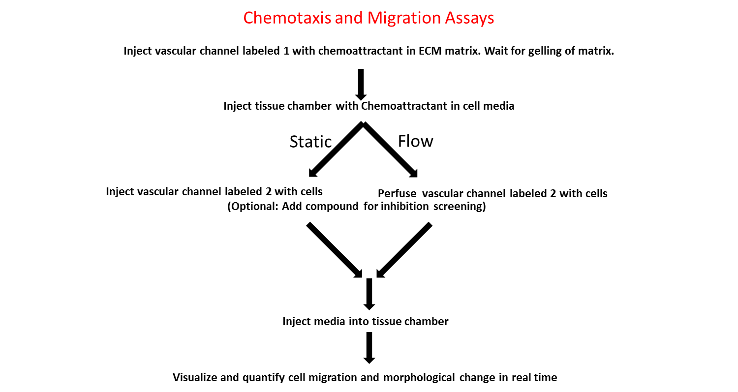
The model has been developed to study the entire inflammation pathway in a realistic and dynamic environment. By creating a cell co-culture and a lumen of endothelial cells, the platform mimics a physiologically realistic model that includes flow and shear. The chip enables real-time tracking of rolling, adhesion and migration processes.
With the Design 1 or 2 - option B (pillar barrier) you can create the inflammation model that provides a realistic testing environment including:
- Physiological shear stress within a microvascular environment
- In vivo like vascular morphology with fully enclosed lumen
- Co-culture capability for cell-cell interactions
- Quantitative real-time rolling, adhesion, and migration data from a single experiment
Inflammation model technical manual
Documentation
?SynBBB technical manual
?SynBBB TEER technical manual
?SynVivo starter kit brochure
?Synvivo brochure
Huang, J., Li, Y. B., Charlebois, C., Nguyen, T., Liu, Z., Bloemberg, D., ... & Jezierski, A. (2022). Application of blood brain barrier models in pre-clinical assessment of glioblastoma-targeting CAR-T based immunotherapies. Fluids and Barriers of the CNS, 19(1), 1-15. https://doi.org/10.1186/s12987-022-00342-y
Brown, T. D., Nowak, M., Bayles, A. V., Prabhakarpandian, B., Karande, P., Lahann, J., ... & Mitragotri, S. (2019). A microfluidic model of human brain (μHuB) for assessment of blood brain barrier. Bioengineering & translational medicine, 4(2), e10126. https://doi.org/10.1002/btm2.10126
Tang, Y., Soroush, F., Sun, S., Liverani, E., Langston, J. C., Yang, Q., ... & Kiani, M. F. (2018). Protein kinase C-delta inhibition protects blood-brain barrier from sepsis-induced vascular damage. Journal of neuroinflammation, 15(1), 1-12. https://doi.org/10.1186/s12974-018-1342-y
Terrell-Hall, T. B., Ammer, A. G., Griffith, J. I., & Lockman, P. R. (2017). Permeability across a novel microfluidic blood-tumor barrier model. Fluids and Barriers of the CNS, 14(1), 1-10. https://doi.org/10.1186/s12987-017-0050-9
Deosarkar, S. P., Prabhakarpandian, B., Wang, B., Sheffield, J. B., Krynska, B., & Kiani, M. F. (2015). A novel dynamic neonatal blood-brain barrier on a chip. PloS one, 10(11), e0142725. https://doi.org/10.1371/journal.pone.0142725
Prabhakarpandian, B., Shen, M. C., Nichols, J. B., Mills, I. R., Sidoryk-Wegrzynowicz, M., Aschner, M., & Pant, K. (2013). SyM-BBB: a microfluidic blood brain barrier model. Lab on a Chip, 13(6), 1093-1101. https://doi.org/10.1039/C2LC41208J